My visit to the Horses Inside Out Anatomy Exhibition
This year as part of the Horses Inside Out conference at Loughborough University in February, there was the opportunity to visit Gillian Higgins’ bigger and more comprehensive than ever Anatomy Exhibition. Either as part of the whole weekend experience, or as a stand alone entry for the exhibition alone.
I had heard a few rumours about some of the additions to this year’s exhibition and was so excited to see it.

The exhibition room was this year going to have 2 additional whole horse skeletons, one of which was fully put together in a canter pose. As well as talk zones with professionals Chris Pearce, Equine Vet & Dental Specialist and Mark Johnson, Farrier & Hoof care Professional, exciting new 3D models painted by Gillian herself and a display of some of Gillian’s incredible art, with the opportunity to purchase some of the pieces to take home!

This was all on top of the previous exhibition pieces some of you may have seen at the conference before, which include the full skeleton of Gillian’s own horse, the original Horses Inside Out horse ‘Freddie Fox’, who was unveiled at the 2023 conference, models of the Sacroiliac region, the hock and the carpus and many other fascinating models both hand made and real bone specimens. And the display of Scientific Posters, this year all related to studies in Growth & Development (the theme of this years conference).
The most recent skeleton addition was a 3 yr & 4 month old Welsh Sec D mare, that had sadly been euthanised following chronic laminitis and had been donated to Gillian for the exhibition. I was fascinated to see this skeleton, firstly as I have a Welsh Sec D myself, and getting the see the size & scale of the bones in a horse similar to my own was incredible. But also to have the opportunity to study the bones and the growth plates, and see just how developed (or not) a horse is at this age, an age at which many of us are starting to ride & train our horses.

Another addition to the exhibition was a foal skeleton, supplied by hoof care practitioner Siobhan Dillon. Again, fascinating to see and compare to the 3yr old mare.
With the 12yr old pony Skeleton and Freddie having been in his 20’s it was possible to see the whole range of bone development as well as bony changes that develop and other pathologies.
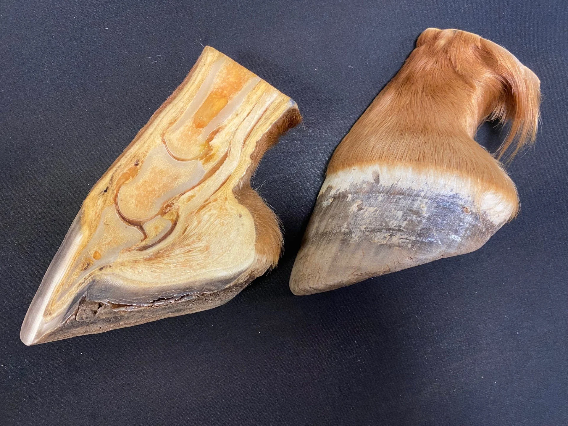
Looking at the feet of the Welsh D was also really interesting, if a little troubling, a stark example of what laminitis can do.

On Mark’s table he had a vast array of preserved legs & feet, showing bone alignment in the leg, pedal bone positioning and what the same foot can look like with and without a balanced trim.
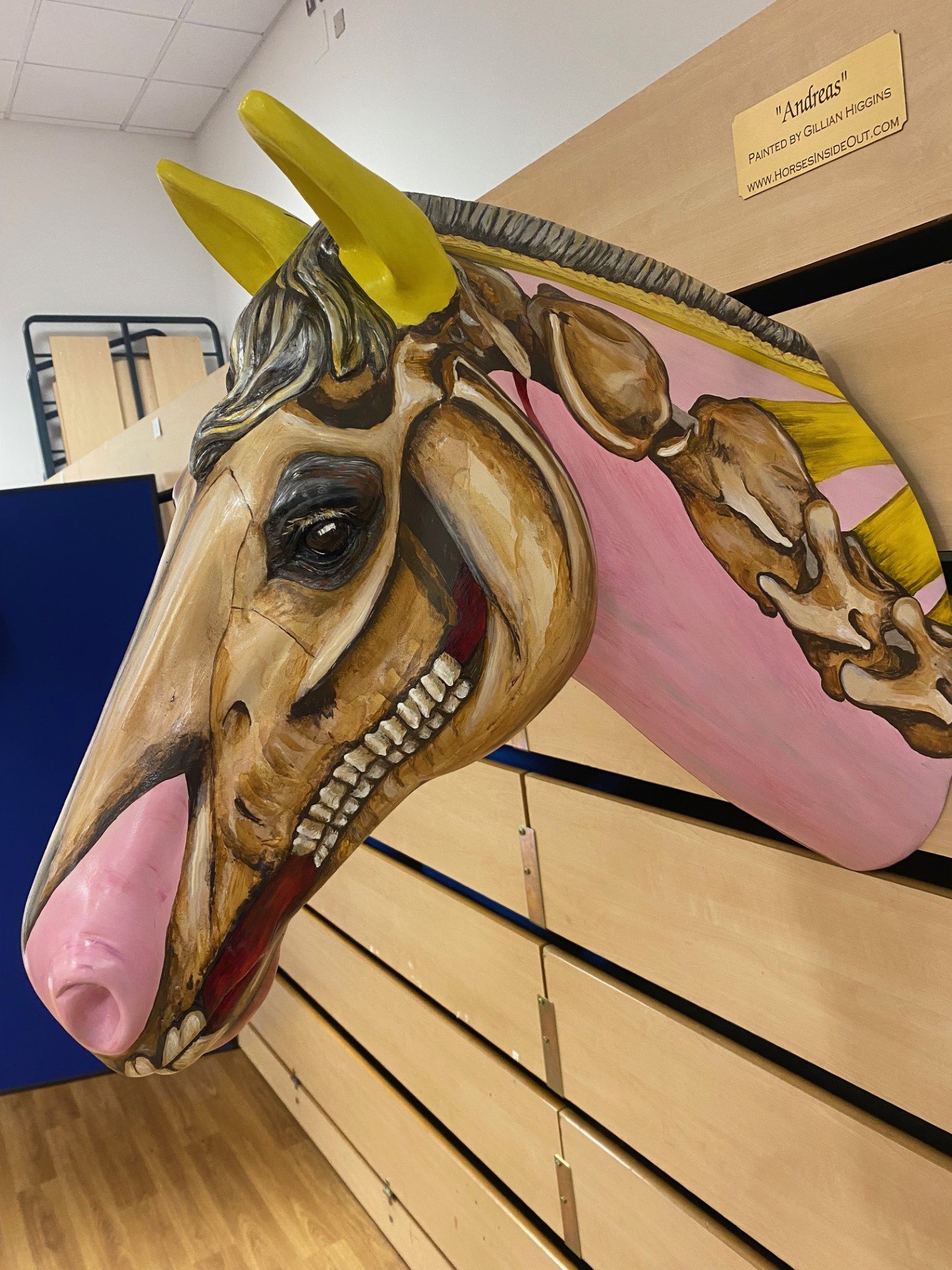
In the head zone was an incredible painted model created by Gillian as well as models of a horses’ teeth at various ages. I really enjoyed studying these.

And I am in awe of some of the art created by Gillian, which is not only beautiful but also insightful and can be used as a learning tool. Some very lucky people got to take home the word art ponies, and statement canvases as well as anatomical posters. What a great fun way to display equine anatomy and spark conversation & intrigue.


There was also the opportunity in the exhibition to label the bones on one of the skeletons and to put a pony skeleton together, as well as build a hock and a carpus joint. You could have spent hours in there being interactive with the bones and the other visitors having discussions. Sadly there just wasn’t enough time to talk about everything!
Luckily there will be another chance at the next conference in 2026 - I am already looking forward to that!
Jess